Achilles Tendon Pain: How a Quick Procedure Helps Patients Get Back on Their Feet
If you have ever dealt with a nagging pain in the back of your heel that won't go away, you might have Achilles tendinopathy. This condition is a common cause of ankle pain, and while physical therapy and exercise are usually the first recommend treatments, they don’t work for everyone. In fact, up to 60% of patients still deal with pain even after trying physical therapy (van der Plas et al., British Journal of Sports Medicine, 2012).
A recent study published in the Journal of Orthopaedic Research investigated a minimally invasive treatment called ultrasound-guided percutaneous tenotomy and debridement to see if it could help patients with Achilles tendinopathy feel better.
What is Achilles Tendinopathy?
The Achilles tendon can be thought of as a heavy-duty bungee cord that connects the calf muscle to the heel bone. It’s the strongest tendon in the body and is used every time we walk, sprint, or jump.
Achilles tendinopathy is what happens when that bungee cord gets "frayed" or irritated because it’s being overloaded. Rather than becoming inflamed, the structure of the Achilles is compromised such that the tendon thickens, tendon fibers are disorganized and often partially torn, and cannot withstand force without producing pain.
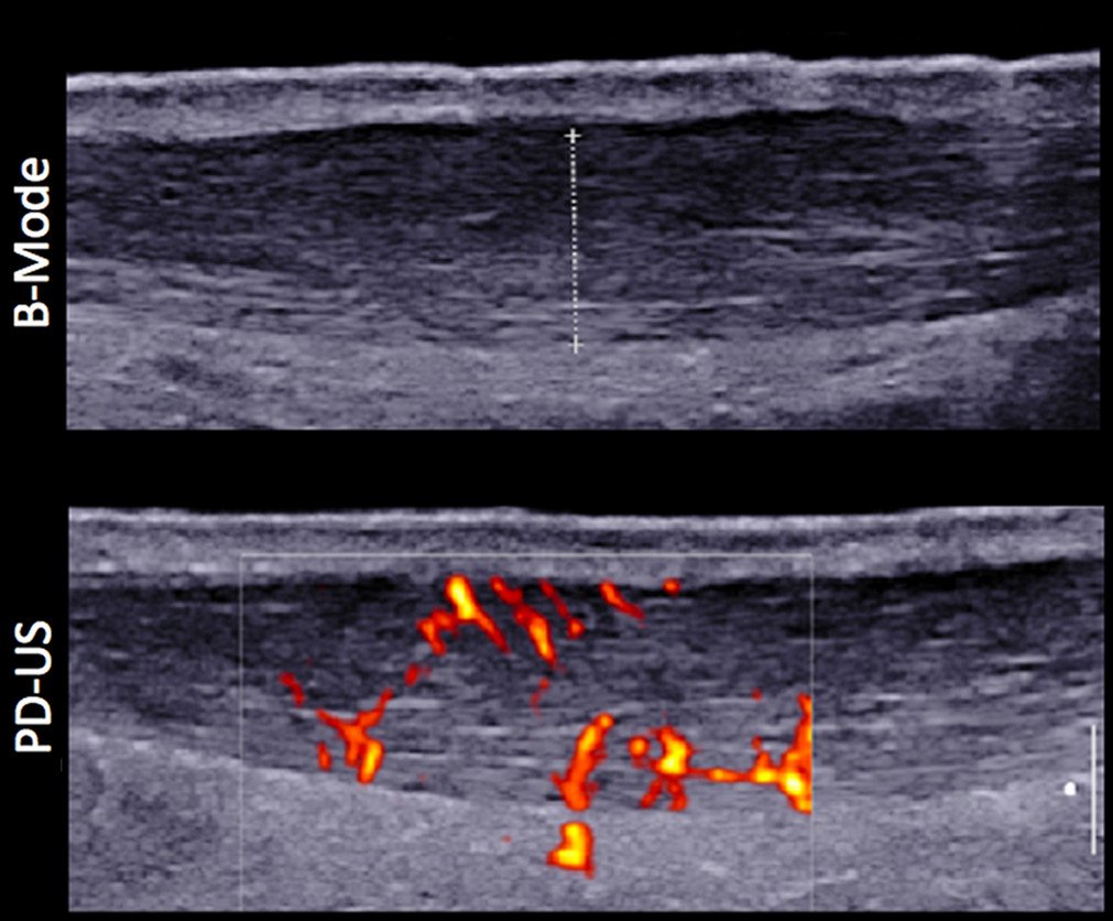
Ultrasound image of mid-portion Achilles tendinopathy (thickened tendon with abnormal fiber architecture with neovascularization). Courtesy of Gatz et al., Orthopaedic Journal of Sports Medicine, 2021
What is the Procedure?
Instead of a major surgery that requires a large incision, ultrasound-guided percutaneous tenotomy and debridement (often referred to as “Tenex”) uses ultrasound imaging to visualize the tendon. A small needle-shaped tool is then used to clean out the damaged parts of the tendon while leaving the healthy parts alone. Because it is minimally invasive, the risk of complications is very low. The incision required to do the procedure is less than one centimeter. The procedure is usually done under local anesthesia and takes less than 10 minutes to perform.
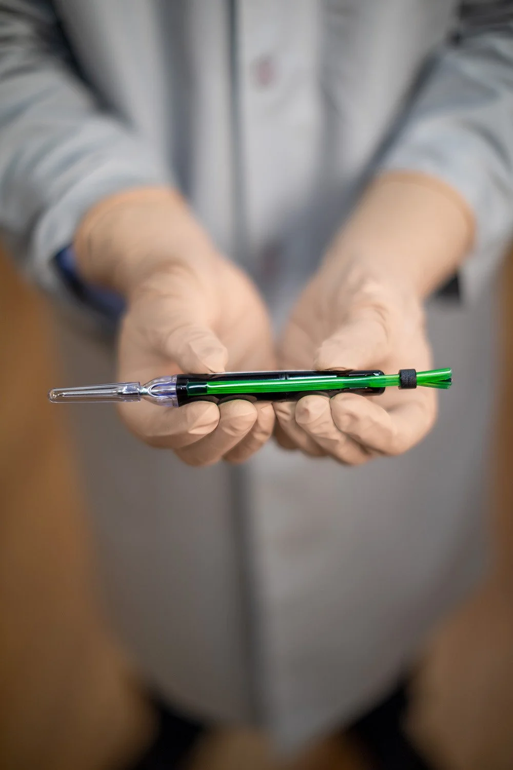
Photo of the Tenex device (Trice Medical), a commercially available tendon debridement tool used to perform ultrasound-guided percutaneous tenotomy and debridement.
What the Study Found
Researchers followed 56 patients for a full year after they had the procedure. The results were encouraging:
The study reported significant improvements across all primary categories:
Pain & Function: There was a "statistically significant and clinically meaningful" improvement in pain scores and functional outcomes as early as 6 weeks post-procedure with results maintained or further improved 1 year after procedure.
Psychological Impact: Interestingly, the study highlighted a reduction in kinesiophobia. As physical pain decreased, patients became significantly less afraid of re-injury during movement.
Safety: No procedure-related complications were reported during the study.
The Bottom Line
For patients who have been struggling with Achilles tendon pain for months and haven't found relief with physical therapy, ultrasound-guided percutaneous needle tenotomy and debridement (“Tenex”) offers a low-risk method to reduce pain, improve function, and get back to an active lifestyle.
Appointment
Dr. Verma provides treatment for numerous orthopaedic and sports medicine conditions of the shoulder, elbow, wrist, hand, hip, knee, ankle and foot. Dr. Verma has specific expertise in ultrasound-guided procedures such as ultrasound-guided percutaneous tenotomy and debridement (“Tenex”) for conditions like Achilles tendinopathy. If you are a patient interested in exploring treatment for your condition, please schedule a consultation with Dr. Verma to discuss the available options.
References
1. Hall, M. M., Chimenti, R. L., Danielson, J. F., & Fleagle, T. R. (2026). Effects of Ultrasound-Guided Tenotomy and Debridement on Pain, Function, and Psychological Factors for Achilles Tendinopathy: A Prospective Cohort Study. Journal of orthopaedic research : official publication of the Orthopaedic Research Society, 44(2), e70071. https://doi.org/10.1002/jor.70071
2. van der Plas, A., de Jonge, S., de Vos, R. J., van der Heide, H. J., Verhaar, J. A., Weir, A., & Tol, J. L. (2012). A 5-year follow-up study of Alfredson's heel-drop exercise programme in chronic midportion Achilles tendinopathy. British journal of sports medicine, 46(3), 214–218. https://doi.org/10.1136/bjsports-2011-090035
3. Gatz, M., Bode, D., Betsch, M., Quack, V., Tingart, M., Kuhl, C., Schrading, S., & Dirrichs, T. (2021). Multimodal Ultrasound Versus MRI for the Diagnosis and Monitoring of Achilles Tendinopathy: A Prospective Longitudinal Study. Orthopaedic journal of sports medicine, 9(4), 23259671211006826. https://doi.org/10.1177/23259671211006826
4. Wong M, Jardaly AH, Kiel J. Anatomy, Bony Pelvis and Lower Limb: Achilles Tendon. [Updated 2023 Aug 8]. In: StatPearls [Internet]. Treasure Island (FL): StatPearls Publishing; 2026 Jan-. Available from: https://www.ncbi.nlm.nih.gov/books/NBK499917/ (NOTE: Blog post thumbnail picture is contributed by OpenStax College, 2017. (Creative Commons Attribution 3.0 license.))